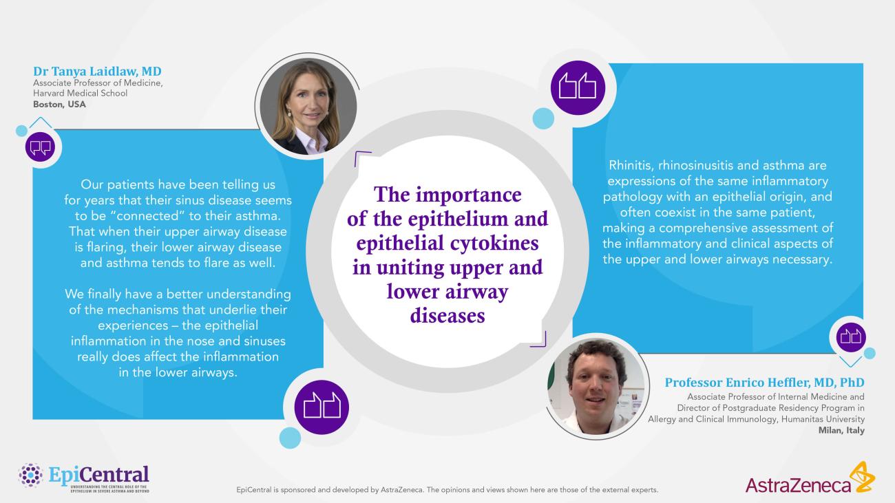
The importance of the epithelium and epithelial cytokines in uniting upper and lower airway diseases

Epithelial dysregulation is implicated in both upper and lower airway diseases.1,2

The frequent co-occurrence of upper and lower airway inflammatory diseases and the immunological links between the two parts of the airways is known as ‘united airways disease’.1,3—5

  • The upper and lower airways are linked anatomically, histologically and immunologically1,3
  • Epithelial barrier dysfunction and epithelial cytokines are linked to chronic inflammatory diseases of multiple organ systems, including lower airway diseases (eg asthma) and upper airway diseases (eg chronic rhinosinusitis and allergic rhinitis)1,2,6,7
  • Upper airway diseases have a negative effect on patients socially and psychologically, severely impacting their quality of life8,9
  • Chronic rhinosinusitis is a heterogeneous inflammatory condition that is characterized by mucosal inflammation of the nasal passage,10,11 which can be divided into two phenotypes based on the presence or absence of nasal polyps11—13
  • Rhinitis is inflammation of the mucous membrane lining in the nasal passages; it can be classified as allergic or non-allergic rhinitis8,14

Understanding the united pathology of upper and lower airway diseases suggests that a united approach to disease management may be needed to achieve global disease control for patients.4,15

References

1. Fokkens W, Reitsma S. Otolaryngol Clin North Am. 2023;56:1–10. 2. Heijink IH, et al. Clin Exp Allergy. 2014;44:620–6303. Jakwerth CA, et al. Cells 2022;11:1387. 4. Kicic A, et al. J Allergy Clin Immunol. 2020;145:1562–1573. 5. Yii AC, et al. Allergy. 2018;73:1964–1978. 6. Bousquet J, et al. Nat Rev Dis Primers. 2020;6:95. 7. Bartemes KR, Kita H. Clin Immunol. 2012;143:222–235. 8. Dykewicz MS, et al. J Allergy Clin Immunol. 2020;146:721–767. 9. Bachert C, et al. J Asthma Allergy. 2021;14:127–134. 10. Lee S, Lane AP. Curr Infect Dis Rep. 2011;13:159–168. 11. Brzost J, et al. Diagnostics (Basel). 2022;12:2301. 12. Orlandi RR, et al. Int Forum Allergy Rhinol. 2016;6(Suppl1):S3–S21. 13. Fokkens WJ, et al. Rhinology. 2020;58(Suppl S29):1–464. 14. Beard S. Prim Care 2014;41:33–46. 15. Licari A, et al. Front Pediatr. 2017;5:44.

Image
Insights from our EpiCollaborators

Insights from our EpiCollaborators

Introduction to the epithelium in the airways 
The airway epithelium is a barrier which maintains homeostasis of the respiratory system and plays an important role in immune function.3,16,17 Epithelial barrier dysfunction is linked to chronic inflammatory diseases of multiple organ systems, including lower airway diseases (eg, asthma) and upper airway diseases (eg, chronic rhinosinusitis and allergic rhinitis).1,2 

The upper and lower airways are linked anatomically, histologically, and immunologically.1,3 Pathological processes which occur in one part of the airway may affect the other; thus, although upper and lower airway diseases can present with different symptoms, the underlying immunological response is often similar.1,4 The frequent co-occurrence of upper and lower airway inflammatory diseases and the immunological links between the two parts of the airways is known as ‘United airways disease’.1,3–5

Image
United airways disease - considering the upper and lower airways as an anatomically and functionally unified organ

'United airways disease' — considering the upper and lower airways as an anatomically and functionally unified organ1,3,15

Upper airway diseases and the role of the epithelium
The nasal airway epithelium is the gateway to the respiratory system, acting as the first point of contact for viruses, pollutants, allergens, and other airborne environmental triggers.18,19 Here, inhaled air is warmed, humidified and filtered, with larger particles trapped by hairs in the nose.1,15,18,20 Smaller particles invade deeper into the airway, where they are captured in secreted mucus and expelled by the action of ciliated cells, in a process known as mucociliary clearance.15,18,20 In addition to the physical barrier provided by the nasal mucosa, in healthy individuals, epithelial cells throughout the airways also initiate an immune response which removes invaders from the respiratory system.3,7,16

Just as epithelial dysregulation is implicated in the pathogenesis of asthma,2,7 disruption of the nasal epithelial barrier contributes to upper airway diseases (eg, allergic rhinitis and chronic rhinosinusitis) through increased permeability and infiltration by external stimuli.1,19 Interaction with external stimuli leads to mucosal inflammation in the upper airways, resulting in the release of cytokines and inflammatory mediators.1,5,20 Inflammation is associated with upper airway remodeling, for example, fibrosis, basement membrane thickening, goblet cell hyperplasia, polyp formation, osteitis, and angiogenesis.13,20 Epithelial cytokines (thymic stromal lymphopoietin [TSLP], interleukin [IL]-33, and IL-25), released by the epithelium in response to triggers, have been implicated in the pathogenesis of upper airway diseases, such as chronic rhinosinusitis and allergic rhinitis.1,6 

Image
Similar inflammatory processes are associated with both upper and lower airways diseases

Similar inflammatory processes are associated with both upper and lower airways diseases1,5,20

Broadly, the airway epithelium consists of a continuous, impermeable sheet of cells, held together with tight junctions and adhesion proteins, which sits on the basement membrane.15,18,19,21,22 In both the upper and lower airways, ciliated and secretory cells facilitate clearance of mucus and airway debris.15,18,20,22 In the upper airways, about 20% of the epithelium is composed of goblet cells,18 which produce mucus with antioxidant, antiprotease, and antimicrobial properties.21 Moving into the lower airways and bronchioles, there are fewer goblet cells18,21; instead, club cells secrete products that have anti-inflammatory and immunosuppressive capacity,18,22 which helps to maintain homeostasis.22 At the end of the bronchial tree, Type 1 alveolar cells, which facilitate gas exchange, and Type 2 alveolar cells, which secrete surfactants, form the alveoli.18 A key factor which differentiates the lower airway epithelium from the upper is the presence of smooth muscle, which drives bronchoconstriction.1,23

Image
Similarities in the cellular composition and structure of the upper and lower airway epithelium
Similarities in the cellular composition and structure of the upper and lower airway epithelium.1,15,20

Chronic rhinosinusitis


Chronic rhinosinusitis is a heterogeneous inflammatory condition that is characterized by mucosal inflammation of the nasal passage.10,11 In Europe, the prevalence is approximately 11%, although this varies widely between countries.13 Chronic rhinosinusitis is divided into two phenotypes based on the presence (CRSwNP) or absence (CRSsNP) of nasal polyps.11–13 The majority (~80%) of patients with chronic rhinosinusitis present without nasal polyps.24,25 

CRSwNP 

Nasal polyps are gray masses within the nose or sinuses that originate from the ethmoid sinus and project into the nasal cavity.16,26 CRSwNP is characterized by nasal congestion, a decreased sense of smell and taste, and sleep disruption.9,13,27 These symptoms place a substantial burden on patients, severely impacting their quality of life.9,27,28 Patients report negative impacts on their psychological and social wellbeing due to decreased enjoyment of food, embarrassment about their symptoms (ie, nasal discharge), and poor sleep quality.9,28

Key unmet needs remain for patients with CRSwNP, including feeling that the burden of disease they experience is underestimated by healthcare providers, and the ineffectiveness of current available treatments.28

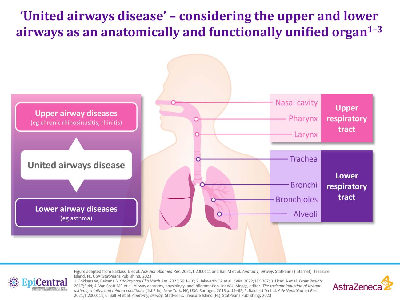
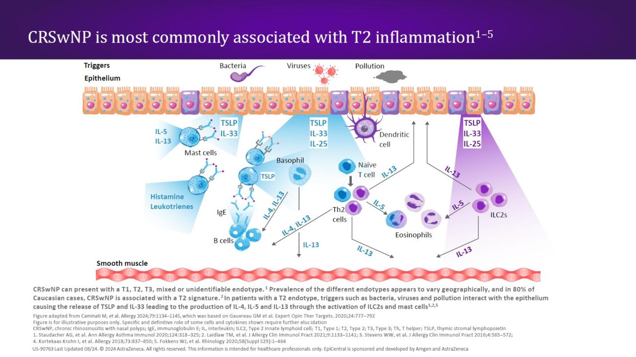
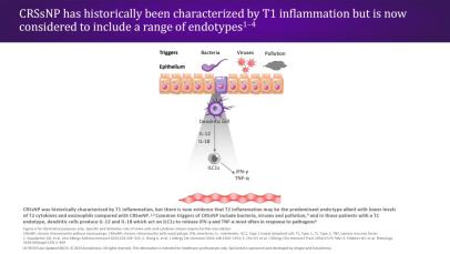
CRSwNP is most commonly associated with a Type 2 (T2) endotype, although many patients show evidence of a mixed endotype.25,29 Endotypes vary geographically24; while T2 inflammation is the predominant endotype in Western countries,25,29 Asian countries may have higher proportions of Type 1 (T1) and Type 3 (T3) disease.24,29 In recent decades, there has been an increase in the proportion of T2 endotypes in Asian countries, which may be caused by increasingly Western lifestyles in this region.24

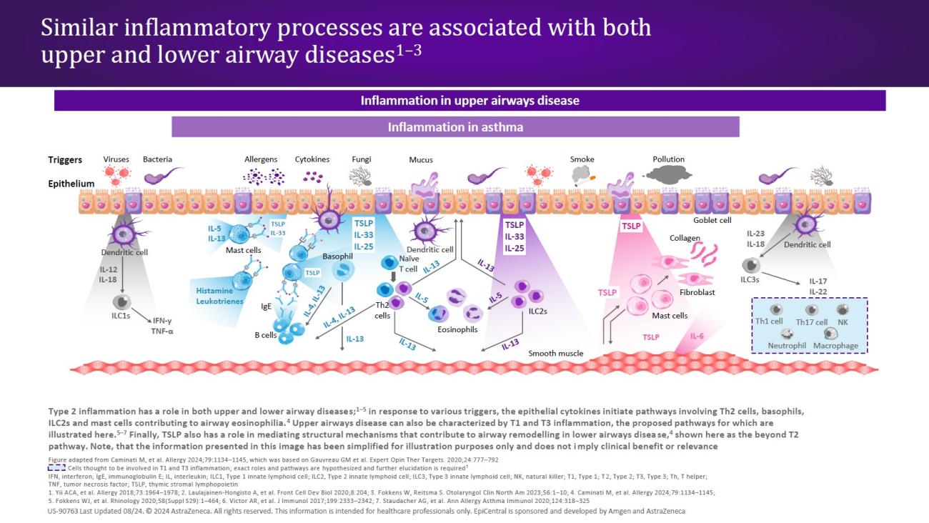
In patients with a T2 endotype, epithelial disruption leads to a cycle of T2 inflammation.30 The defective epithelial barrier is more easily permeable, and epithelial cytokines (TSLP, IL-33, and IL-25) are released in response to external triggers acting on the epithelium.16,30 All three epithelial cytokines have been shown to be overexpressed in patients with CRSwNP.31–33 The release of TSLP and IL-33 in turn leads to the production of downstream mediators (ie, IL-4, IL-5, and IL-13) through the activation of innate Type 2 lymphoid cells (ILC2s) and mast cells24,30,33; ILC2s, mast cells, and the T2 cytokines IL-5 and IL-13 have all shown to be elevated in patients with CRSwNP.16 These cytokines amplify the T2 inflammatory response,30,33 while IL-4 and IL-13 may perpetuate barrier dysfunction through induction of additional TSLP expression.30

Image
Similar inflammatory processes are associated with both upper and lower airways diseases
In patients with a T2 endotype, the release of TSLP and IL-33 leads to the production of IL-4, IL-5, and IL-13 through the activation of ILC2s and mast cells.24,30,33

CRSsNP 

Of the two phenotypes, CRSsNP is considered to be the less severe disease.16,24 Patients experience the nasal congestion and altered smell and taste that is characteristic of CRSwNP; additionally, nasal discharge and facial pain/pressure are key symptoms for patients with CRSsNP.13,25  

For patients with CRSsNP, a key unmet need is the limited efficacy evidence for some existing treatments, in addition to the lack of research into novel treatments, as compared with CRSwNP.12,24,34 

CRSsNP is a heterogeneous disease, characterized by multiple inflammatory endotypes.25,34 Previously, CRSsNP was associated with T1 inflammation24; however, it is now clear that the picture is more complex.25 As with CRSwNP (albeit to a lesser extent), T2 inflammation is the dominant endotype for CRSsNP in Western patients,24,25 whereas in some Asian populations, T1 inflammation predominates.24 The presence of overlapping endotypes was found to be common (26%) in a study of US patients, as was the presence of no identifiable endotype (30%).25

In patients with a T1 endotype, external stimuli acting on the epithelium activate dendritic cells (DCs) to produce IL-12 and IL-18.13 These act on innate Type 1 lymphoid cells (ILC1s) to release interferon (IFN)-γ and tumor necrosis factor (TNF)-α.13 It has been suggested that bacteria may play a role in CRSsNP35; however, reports are conflicting and further research is needed to understand causality.35,36 

Image
Similar inflammatory processes are associated with both upper and lower airways diseases
In patients with a T1 endotype, DCs produce IL-12 and IL-18 which act on ILC1s to release IFN-γ and TNF-α.13

Nasal irrigation with saline solution, alongside topical corticosteroids, are recommended treatments for CRSwNP and CRSsNP.12,34 Oral corticosteroids (OCS) are used as an additional short-term treatment to reduce polyp size for patients with CRSwNP.9,12,37 Patients with CRSsNP are also treated with OCS34; however, this is not recommended owing to the lack of efficacy data.12 In both diseases the risk of adverse side effects associated with OCS use (gastrointestinal symptoms, insomnia, osteoporosis)13,37 must be balanced with the potential benefit of treatment.12 If these treatments are ineffective then endoscopic sinus surgery may be indicated12,34,38; however, the benefits are often short-lived,34,38 with revision rates of ~30% in CRSwNP and ~10% in CRSsNP.39 Furthermore, the time between surgeries decreases with each subsequent revision.39

Rhinitis

Rhinitis is inflammation of the mucous membrane lining in the nasal passages; it can be classified as allergic or non-allergic rhinitis.8,14 Rhinitis is characterized by the acute or chronic intermittent or persistent presence of one or more nasal symptoms (also known as nasal hyperreactivity),40 which include sneezing, nasal blockage or congestion, itching, and nasal discharge.6,15,40,41   

Globally, the median prevalence for rhinitis is ~29%, with median prevalence of allergic rhinitis and non-allergic rhinitis being ~18% and 12%, respectively; this varies according to geographic location.42 Patients with rhinitis typically experience an impaired quality of life, with sleep, exercise tolerance, and physical and social function all impacted8,43; the economic and societal burden of rhinitis is also high.8 

Allergic rhinitis

Allergic rhinitis occurs following an immunoglobulin E (IgE)-mediated reaction to inhaled allergens, resulting in T2 inflammation in the respiratory tract.5 There remains an unmet need in the diagnosis of allergic rhinitis, owing to poor public awareness, limited access to specialists (eg, allergologists), and confounding diagnoses.6,44

The early allergic response in allergic rhinitis involves allergen binding to IgE, triggering mast cell degranulation, resulting in acute allergic symptoms.5,41 The late allergic response involves epithelial cytokines TSLP, IL-33, and IL-25 being released from the epithelium during allergen exposure.5,6,43 These cytokines play a role in the initiation and maintenance of T2 inflammation through production of cytokines IL-4, IL-5, and IL-13,5,6,41,43 all of which can lead to chronic allergic symptoms and inflammation in allergic rhinitis, as well as remodeling and nasal hyperresponsiveness.5,41 Allergic rhinitis is also associated with an increased onset of airway hyperresponsiveness.45,46 

Image
Similar inflammatory processes are associated with both upper and lower airways diseases
In patients with allergic rhinitis, TSLP, IL-33, and IL-25 are released following allergen exposure, leading to T2 inflammation through the cytokines IL-4, IL-5, and IL-13.5,41,43

Learn more about the pathophysiology and clinical significance of airway hyperresponsiveness and airway remodeling.

Non-allergic rhinitis  

Non-allergic rhinitis includes a group of heterogeneous rhinitis conditions, independent of an IgE-mediated mechanism and characterized by the presence of at least two nasal symptoms.15,41,47 Patients with non-allergic rhinitis often have similar symptoms to patients with allergic rhinitis, but experience less sneezing and itching and more nasal congestion, nasal discharge, and sinus headaches.48 

Non-allergic rhinitis is also associated with significant disease burden8,40; improvements in understanding of this disease and development of novel assessment and diagnostic tools are needed.40

The exact mechanism of non-allergic rhinitis is still poorly understood, owing to the multiple heterogeneous presentations of the disease and a lack of both a uniform definition and international consensus on diagnostic criteria.14,40,41 However, ILC2s are hypothesized to be potentially involved in a non-allergic rhinitis subtype (non-allergic rhinitis with eosinophilia syndrome [NARES]).5 In this process, TSLP, IL-33, and IL-25 can activate ILC2s, leading to eosinophil activation via production of IL-5.5 Infiltration of mast cells into nasal tissue has been implicated in other subtypes, non-allergic rhinitis eosinophilic mast cell syndrome (NARESMA), and non-allergic rhinitis with mast cells (NARMA).49

Image
Similar inflammatory processes are associated with both upper and lower airways diseases
In patients with NARES, it is hypothesized that TSLP, IL-33, and IL-25 activate ILC2s, which leads to eosinophil activation via IL-5.5

United airways disease 

United airways disease proposes that the upper and lower airways form a single organ, whereby a local pathological response would impact the whole respiratory tract.4,5 Under this theory, diseases of the upper and lower airways, which appear distinct, may reflect local manifestations of a wider, systemic immune response.5,15 This supports the finding that upper and lower airway diseases are frequently comorbid.1,50 Understanding the united pathology of these diseases suggests that a united approach to disease management may be needed to achieve global disease control for patients.4,15 

What percentage of patients have comorbid upper and lower airways disease?

  • 20 to 25% of patients with chronic rhinosinusitis also have asthma50
  • 5 to 10% of patients with asthma have comorbid chronic rhinosinusitis50 
  • 19 to 38% of patients with allergic rhinitis also have asthma15
  • 30 to 80% of patients with asthma have comorbid allergic rhinitis15

Diagnostic tools and biomarkers in upper airway diseases

Currently available biomarkers for upper airway diseases include blood eosinophils, total serum IgE, and allergen-specific IgE from a blood or skin-prick test.6,13,51 Such biomarkers may demonstrate the presence of T2 disease; however, their clinical utility beyond this remains unclear.6,13 Other potentially promising techniques include examination of tissue biopsies, determination of cytokine levels in nasal lavage fluid, or nasal cytology.6,49,51 Nasal cytology provides an insight into the inflammatory cells infiltrating the nasal mucosa, which has allowed for the identification of specific pathologies in rhinitis and chronic rhinosinusitis.52 It is hoped that through further research, in the future, biomarkers and other diagnostic tools, which allow for upper airway disease to be accurately subtyped, will lead to personalized treatment and better outcomes for patients.13,51 

The content for this module was created with the support of Dr Tanya Laidlaw and Professor Enrico Heffler.

RELATED RESOURCES

Like this page? We have a host of other resources available in the ‘Scientific and Resource Library’ where you can find out more.

References 

1. Fokkens W, Reitsma S. Otolaryngol Clin North Am. 2023;56:1–10. 2. Heijink IH, et al. Clin Exp Allergy. 2014;44:620–630. 3. Jakwerth CA, et al. Cells. 2022;11:1387. 4. Kicic A, et al. J Allergy Clin Immunol. 2020;145:1562–1573. 5. Yii AC, et al. Allergy. 2018;73:1964–1978. 6. Bousquet J, et al. Nat Rev Dis Primers. 2020;6:95. 7. Bartemes KR, Kita H. Clin Immunol. 2012;143:222–235. 8. Dykewicz MS, et al. J Allergy Clin Immunol. 2020;146:721–767. 9. Bachert C, et al. J Asthma Allergy. 2021;14:127–134. 10. Lee S, Lane AP. Curr Infect Dis Rep. 2011;13:159–168. 11. Brzost J, et al. Diagnostics (Basel). 2022;12:2301. 12. Orlandi RR, et al. Int Forum Allergy Rhinol. 2016;6(Suppl 1):S3–S21. 13. Fokkens WJ, et al. Rhinology. 2020;58(Suppl S29):1–464. 14. Beard S. Prim Care. 2014;41:33–46. 15. Licari A, et al. Front Pediatr. 2017;5:44. 16. Stevens WW, et al. J Allergy Clin Immunol Pract. 2016;4:565–572. 17. Roan F, et al. J Clin Invest. 2019;129:1441–1451. 18. Adivitiya, et al. Biology (Basel). 2021;10:95. 19. Zhang R, et al. Int Arch Allergy Immunol. 2023;184:1–21. 20. Laulajainen-Hongisto A, et al. Front Cell Dev Biol. 2020;8:204. 21. Crystal RG, et al. Proc Am Thorac Soc. 2008;5:772–777. 22. Davis JD, Wypych TP. Mucosal Immunol. 2021;14:978–990. 23. Doeing DC, Solway J. J Appl Physiol (1985). 2013;114:834–843. 24. Staudacher AG, et al. Ann Allergy Asthma Immunol. 2020;124:318–325. 25. Stevens WW, et al. J Allergy Clin Immunol Pract. 2019;7:2812–2820.e3. 26. Newton JR, Ah-See KW. Ther Clin Risk Manag. 2008;4:507–512. 27. Chen SY, et al. Rhinol Online. 2022;5:157–173. 28. Claeys N, et al. Front Allergy. 2021;2:761388. 29. Hao D, et al. J Inflamm Res. 2022;15:5557–5565. 30. Laidlaw TM, et al. J Allergy Clin Immunol Pract. 2021;9:1133–1141. 31. Deng H, et al. J Asthma Allergy. 2021;14:839–850. 32. Liu R, et al. Front Immunol. 2021;12:530488. 33. Sehmi R. Thorax. 2017;72:591–593. 34. Cho SH, et al. J Allergy Clin Immunol Pract. 2016;4:575–582. 35. Koeller K, et al. Front Microbiol. 2018;9:643. 36. Ramakrishnan VR, et al. J Allergy Clin Immunol. 2015;136:334–342.e1. 37. Head K, et al. Cochrane Database Syst Rev. 2016;4:CD011991. 38. Peters AT, et al. Allergy Asthma Proc. 2022;43:435–445. 39. Smith KA, et al. Int Forum Allergy Rhinol. 2019;9:402–408. 40. Hellings PW, et al. Allergy. 2017;72:1657–1665. 41. Sin B, Togias A. Proc Am Thorac Soc. 2011;8:106–114. 42. Savouré M, et al. Clin Transl Allergy. 2022;12:e12130. 43. Wise SK, et al. Int Forum Allergy Rhinol. 2018;8:108–352. 44. Small P, et al. Allergy Asthma Clin Immunol. 2018;14:51. 45. Liu Y, et al. J Immunol Res. 2022;2022:4351345. 46. Shaaban R, et al. Am J Respir Crit Care Med. 2007;176:659–666. 47. Kaliner MA. World Allergy Organ J. 2009;2:98–101. 48. Greiwe JC, Bernstein JA. J Clin Med. 2019;8:2019. 49. Heffler E, et al. Clin Exp Allergy. 2018;48:1092–1106. 50. Shamil E, Hopkins C. Otolaryngol Clin North Am. 2023;56:157–168. 51. Miglani A, et al. Otolaryngol Clin North Am. 2023;56:11–22. 52. Fokkens WJ, et al. Rhinology. 2020;58(Suppl S29):1–464. 53. Caruso C et al. Front Allergy. 2022;3:768408.